AKR1B1
Protein-coding gene in the species Homo sapiens
| AKR1B1 | |||||||||||||||||||||||||||||||||||||||||||||||||||
|---|---|---|---|---|---|---|---|---|---|---|---|---|---|---|---|---|---|---|---|---|---|---|---|---|---|---|---|---|---|---|---|---|---|---|---|---|---|---|---|---|---|---|---|---|---|---|---|---|---|---|---|
 | |||||||||||||||||||||||||||||||||||||||||||||||||||
| Identifiers | |||||||||||||||||||||||||||||||||||||||||||||||||||
| Aliases | AKR1B1, ADR, ALDR1, ALR2, AR, aldo-keto reductase family 1, member B1 (aldose reductase), aldo-keto reductase family 1 member B | ||||||||||||||||||||||||||||||||||||||||||||||||||
| External IDs | OMIM: 103880; MGI: 1353494; HomoloGene: 133743; GeneCards: AKR1B1; OMA:AKR1B1 - orthologs | ||||||||||||||||||||||||||||||||||||||||||||||||||
| |||||||||||||||||||||||||||||||||||||||||||||||||||
| |||||||||||||||||||||||||||||||||||||||||||||||||||
| |||||||||||||||||||||||||||||||||||||||||||||||||||
| |||||||||||||||||||||||||||||||||||||||||||||||||||
| |||||||||||||||||||||||||||||||||||||||||||||||||||
| Wikidata | |||||||||||||||||||||||||||||||||||||||||||||||||||
| |||||||||||||||||||||||||||||||||||||||||||||||||||


Aldo-keto reductase family 1, member B1 (AKR1B1), also known as aldose reductase, is an enzyme that is encoded by the AKR1B1 gene in humans.[5][6] It is a reduced nicotinamide-adenine dinucleotide phosphate (NADPH)-dependent enzyme catalyzing the reduction of various aldehydes and ketones to the corresponding alcohol. The involvement of AKR1B1 in oxidative stress diseases, cell signal transduction, and cell proliferation process endows AKR1B1 with potential as a therapeutic target.
Structure
Gene
The AKR1B1 gene lies on the chromosome location of 7q33 and consists of 10 exons. There are a few putative pseudogenes for this gene, and one of them has been confirmed and mapped to chromosome 3.[6]
Protein
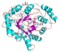
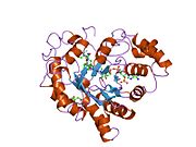
AKR1B1 consists of 316 amino acid residues and weighs 35853Da. It does not possess the traditional dinucleotide binding fold. The way it binds NADPH differs from other nucleotide adenine dinucleotide-dependent enzymes. The active site pocket of human aldose reductase is relatively hydrophobic, lined by seven aromatic and four other non-polar residues.[7]
Function
AR belongs to the aldehyde-keto reductase superfamily, with a widely expression in human organs including the kidney, lens, retina, nerve, heart, placenta, brain, skeletal muscle, testis, blood vessels, lung, and liver.[8] It is a reduced nicotinamide-adenine dinucleotide phosphate (NADPH)-dependent enzyme catalyzing the reduction of various aldehydes and ketones to the corresponding alcohol. It also participates in glucose metabolism and osmoregulation and plays a protective role against toxic aldehydes derived from lipid peroxidation and steroidogenesis.[9]
Clinical significance
Under diabetic conditions AR converts glucose into sorbitol, which is then converted to fructose. 20466987 It has been found to play an important role in many diabetes complications such as diabetes retinopathy and renopathy.[10][11][12] It is also involved in many oxidative stress diseases, cell signal transduction, and cell proliferation process including cardiovascular disorders, sepsis, and cancer.[13]
It has been reported that the action of AR contributes to the activation of retinal microglia, suggesting that inhibition of AR may be of a therapeutic importance to reduce inflammation associated with activation of RMG.[14] Adapting AR inhibitors could as well prevent sepsis complications, prevent angiogenesis, ameliorate mild or asymptomatic diabetic cardiovascular autonomic neuropathy and may be a promising strategy for the treatment of endotoxemia and other ROS-induced inflammatory diseases.[12]
Interactions
AKR1B1 has been found to interact with:
- ginsenoside 20(S)-Rh2[15]
- alkaloid[16]
- carboxylic acid derivatives[12]
- spirohydantoins[12]
- cyclic amides[12]
References
- ^ a b c GRCh38: Ensembl release 89: ENSG00000085662 – Ensembl, May 2017
- ^ a b c GRCm38: Ensembl release 89: ENSMUSG00000001642 – Ensembl, May 2017
- ^ "Human PubMed Reference:". National Center for Biotechnology Information, U.S. National Library of Medicine.
- ^ "Mouse PubMed Reference:". National Center for Biotechnology Information, U.S. National Library of Medicine.
- ^ Graham A, Heath P, Morten JE, Markham AF (March 1991). "The human aldose reductase gene maps to chromosome region 7q35". Human Genetics. 86 (5): 509–14. doi:10.1007/BF00194644. PMID 1901827. S2CID 34446965.
- ^ a b "Entrez Gene: AKR1B1 aldo-keto reductase family 1, member B1 (aldose reductase)".
- ^ Lee H (August 1998). "The structure and function of yeast xylose (aldose) reductases". Yeast. 14 (11): 977–84. doi:10.1002/(sici)1097-0061(199808)14:11<977::aid-yea302>3.0.co;2-j. PMID 9730277. S2CID 39792612.
- ^ O'connor T, Ireland LS, Harrison DJ, Hayes JD (October 1999). "Major differences exist in the function and tissue-specific expression of human aflatoxin B1 aldehyde reductase and the principal human aldo-keto reductase AKR1 family members". The Biochemical Journal. 343 Pt 2 (2): 487–504. doi:10.1042/bj3430487. PMC 1220579. PMID 10510318.
- ^ Lefrançois-Martinez AM, Bertherat J, Val P, Tournaire C, Gallo-Payet N, Hyndman D, Veyssière G, Bertagna X, Jean C, Martinez A (June 2004). "Decreased expression of cyclic adenosine monophosphate-regulated aldose reductase (AKR1B1) is associated with malignancy in human sporadic adrenocortical tumors". The Journal of Clinical Endocrinology and Metabolism. 89 (6): 3010–9. doi:10.1210/jc.2003-031830. PMID 15181092.
- ^ Park J, Kim H, Park SY, Lim SW, Kim YS, Lee DH, Roh GS, Kim HJ, Kang SS, Cho GJ, Jeong BY, Kwon HM, Choi WS (May 2014). "Tonicity-responsive enhancer binding protein regulates the expression of aldose reductase and protein kinase C δ in a mouse model of diabetic retinopathy". Experimental Eye Research. 122: 13–9. doi:10.1016/j.exer.2014.03.001. PMID 24631337.
- ^ Zhou M, Zhang P, Xu X, Sun X (April 2015). "The Relationship Between Aldose Reductase C106T Polymorphism and Diabetic Retinopathy: An Updated Meta-Analysis". Investigative Ophthalmology & Visual Science. 56 (4): 2279–89. doi:10.1167/iovs.14-16279. PMID 25722213.
- ^ a b c d e Grewal AS, Bhardwaj S, Pandita D, Lather V, Sekhon BS (2016-01-01). "Updates on Aldose Reductase Inhibitors for Management of Diabetic Complications and Non-diabetic Diseases". Mini Reviews in Medicinal Chemistry. 16 (2): 120–62. doi:10.2174/1389557515666150909143737. PMID 26349493.
- ^ Maccari R, Ottanà R (March 2015). "Targeting aldose reductase for the treatment of diabetes complications and inflammatory diseases: new insights and future directions". Journal of Medicinal Chemistry. 58 (5): 2047–67. doi:10.1021/jm500907a. PMID 25375908.
- ^ Chang KC, Ponder J, Labarbera DV, Petrash JM (May 2014). "Aldose reductase inhibition prevents endotoxin-induced inflammatory responses in retinal microglia". Investigative Ophthalmology & Visual Science. 55 (5): 2853–61. doi:10.1167/iovs.13-13487. PMC 4010364. PMID 24677107.
- ^ Fatmawati S, Ersam T, Yu H, Zhang C, Jin F, Shimizu K (September 2014). "20(S)-Ginsenoside Rh2 as aldose reductase inhibitor from Panax ginseng". Bioorganic & Medicinal Chemistry Letters. 24 (18): 4407–9. doi:10.1016/j.bmcl.2014.08.009. PMID 25152999.
- ^ Gupta S, Singh N, Jaggi AS (March 2014). "Alkaloids as aldose reductase inhibitors, with special reference to berberine". Journal of Alternative and Complementary Medicine. 20 (3): 195–205. doi:10.1089/acm.2013.0088. PMID 24236461.
Further reading
- Borhani DW, Harter TM, Petrash JM (December 1992). "The crystal structure of the aldose reductase.NADPH binary complex". The Journal of Biological Chemistry. 267 (34): 24841–7. doi:10.2210/pdb1abn/pdb. PMID 1447221.
- Wilson DK, Bohren KM, Gabbay KH, Quiocho FA (July 1992). "An unlikely sugar substrate site in the 1.65 A structure of the human aldose reductase holoenzyme implicated in diabetic complications". Science. 257 (5066): 81–4. doi:10.1126/science.1621098. PMID 1621098.
- Graham A, Heath P, Morten JE, Markham AF (March 1991). "The human aldose reductase gene maps to chromosome region 7q35". Human Genetics. 86 (5): 509–14. doi:10.1007/BF00194644. PMID 1901827. S2CID 34446965.
- Graham A, Brown L, Hedge PJ, Gammack AJ, Markham AF (April 1991). "Structure of the human aldose reductase gene". The Journal of Biological Chemistry. 266 (11): 6872–7. doi:10.1016/S0021-9258(20)89582-9. PMID 1901857.
- Grundmann U, Bohn H, Obermeier R, Amann E (April 1990). "Cloning and prokaryotic expression of a biologically active human placental aldose reductase". DNA and Cell Biology. 9 (3): 149–57. doi:10.1089/dna.1990.9.149. PMID 2111143.
- Nishimura C, Matsuura Y, Kokai Y, Akera T, Carper D, Morjana N, Lyons C, Flynn TG (June 1990). "Cloning and expression of human aldose reductase". The Journal of Biological Chemistry. 265 (17): 9788–92. doi:10.1016/S0021-9258(19)38740-X. PMID 2112546.
- Morjana NA, Lyons C, Flynn TG (February 1989). "Aldose reductase from human psoas muscle. Affinity labeling of an active site lysine by pyridoxal 5'-phosphate and pyridoxal 5'-diphospho-5'-adenosine". The Journal of Biological Chemistry. 264 (5): 2912–9. doi:10.1016/S0021-9258(19)81699-X. PMID 2492527.
- Bohren KM, Bullock B, Wermuth B, Gabbay KH (June 1989). "The aldo-keto reductase superfamily. cDNAs and deduced amino acid sequences of human aldehyde and aldose reductases". The Journal of Biological Chemistry. 264 (16): 9547–51. doi:10.1016/S0021-9258(18)60566-6. PMID 2498333.
- Chung S, LaMendola J (September 1989). "Cloning and sequence determination of human placental aldose reductase gene". The Journal of Biological Chemistry. 264 (25): 14775–7. doi:10.1016/S0021-9258(18)63766-4. PMID 2504709.
- Graham A, Hedge PJ, Powell SJ, Riley J, Brown L, Gammack A, Carey F, Markham AF (October 1989). "Nucleotide sequence of cDNA for human aldose reductase". Nucleic Acids Research. 17 (20): 8368. doi:10.1093/nar/17.20.8368. PMC 334974. PMID 2510130.
- Akagi Y, Kador PF, Kuwabara T, Kinoshita JH (November 1983). "Aldose reductase localization in human retinal mural cells". Investigative Ophthalmology & Visual Science. 24 (11): 1516–9. PMID 6417042.
- Ko BC, Lam KS, Wat NM, Chung SS (July 1995). "An (A-C)n dinucleotide repeat polymorphic marker at the 5' end of the aldose reductase gene is associated with early-onset diabetic retinopathy in NIDDM patients". Diabetes. 44 (7): 727–32. doi:10.2337/diabetes.44.7.727. PMID 7789640.
- Wilson DK, Tarle I, Petrash JM, Quiocho FA (November 1993). "Refined 1.8 A structure of human aldose reductase complexed with the potent inhibitor zopolrestat". Proceedings of the National Academy of Sciences of the United States of America. 90 (21): 9847–51. Bibcode:1993PNAS...90.9847W. doi:10.1073/pnas.90.21.9847. PMC 47669. PMID 8234324.
- Tarle I, Borhani DW, Wilson DK, Quiocho FA, Petrash JM (December 1993). "Probing the active site of human aldose reductase. Site-directed mutagenesis of Asp-43, Tyr-48, Lys-77, and His-110". The Journal of Biological Chemistry. 268 (34): 25687–93. doi:10.1016/S0021-9258(19)74444-5. PMID 8245005.
- Robinson B, Hunsaker LA, Stangebye LA, Vander Jagt DL (December 1993). "Aldose and aldehyde reductases from human kidney cortex and medulla". Biochimica et Biophysica Acta (BBA) - Protein Structure and Molecular Enzymology. 1203 (2): 260–6. doi:10.1016/0167-4838(93)90092-6. PMID 8268209.
- Jaquinod M, Potier N, Klarskov K, Reymann JM, Sorokine O, Kieffer S, Barth P, Andriantomanga V, Biellmann JF, Van Dorsselaer A (December 1993). "Sequence of pig lens aldose reductase and electrospray mass spectrometry of non-covalent and covalent complexes". European Journal of Biochemistry. 218 (3): 893–903. doi:10.1111/j.1432-1033.1993.tb18445.x. PMID 8281941.
- Liu SQ, Bhatnagar A, Ansari NH, Srivastava SK (August 1993). "Identification of the reactive cysteine residue in human placenta aldose reductase". Biochimica et Biophysica Acta (BBA) - Protein Structure and Molecular Enzymology. 1164 (3): 268–72. doi:10.1016/0167-4838(93)90258-S. PMID 8343525.
- Nishimura C, Furue M, Ito T, Omori Y, Tanimoto T (July 1993). "Quantitative determination of human aldose reductase by enzyme-linked immunosorbent assay. Immunoassay of human aldose reductase". Biochemical Pharmacology. 46 (1): 21–8. doi:10.1016/0006-2952(93)90343-U. PMID 8347133.
- Sato S, Lin LR, Reddy VN, Kador PF (August 1993). "Aldose reductase in human retinal pigment epithelial cells". Experimental Eye Research. 57 (2): 235–41. doi:10.1006/exer.1993.1119. PMID 8405190.
- Ferraretto A, Negri A, Giuliani A, De Grada L, Fuhrman Conti AM, Ronchi S (February 1993). "Aldose reductase is involved in long-term adaptation of EUE cells to hyperosmotic stress". Biochimica et Biophysica Acta (BBA) - Molecular Cell Research. 1175 (3): 283–8. doi:10.1016/0167-4889(93)90218-E. PMID 8435445.
External links
- Human AKR1B1 genome location and AKR1B1 gene details page in the UCSC Genome Browser.
- Human AR genome location and AR gene details page in the UCSC Genome Browser.
- v
- t
- e
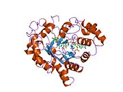
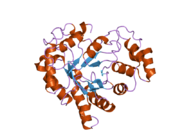
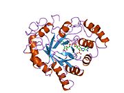
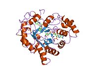
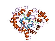
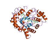
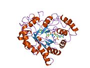
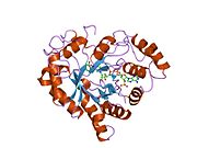
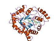
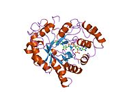
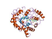
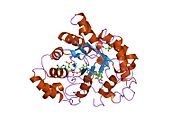
PDB gallery
-
 1abn: THE CRYSTAL STRUCTURE OF THE ALDOSE REDUCTASE NADPH BINARY COMPLEX
 -
 1ads: AN UNLIKELY SUGAR SUBSTRATE SITE IN THE 1.65 ANGSTROMS STRUCTURE OF THE HUMAN ALDOSE REDUCTASE HOLOENZYME IMPLICATED IN DIABETIC COMPLICATIONS
 -
 1az1: ALRESTATIN BOUND TO C298A/W219Y MUTANT HUMAN ALDOSE REDUCTASE
 -
 1az2: CITRATE BOUND, C298A/W219Y MUTANT HUMAN ALDOSE REDUCTASE
 -
 1ef3: FIDARESTAT BOUND TO HUMAN ALDOSE REDUCTASE
 -
 1el3: HUMAN ALDOSE REDUCTASE COMPLEXED WITH IDD384 INHIBITOR
 -
 1iei: CRYSTAL STRUCTURE OF HUMAN ALDOSE REDUCTASE COMPLEXED WITH THE INHIBITOR ZENARESTAT.
 -
 1mar: REFINED 1.8 ANGSTROMS STRUCTURE OF HUMAN ALDOSE REDUCTASE COMPLEXED WITH THE POTENT INHIBITOR ZOPOLRESTAT
 -
 1pwl: Crystal structure of human Aldose Reductase complexed with NADP and Minalrestat
 -
 1pwm: Crystal structure of human Aldose Reductase complexed with NADP and Fidarestat
 -
 1t40: Crystal structure of human aldose reductase complexed with NADP and IDD552 at ph 5
 -
 1t41: Crystal structure of human aldose reductase complexed with NADP and IDD552
 -
 1us0: HUMAN ALDOSE REDUCTASE IN COMPLEX WITH NADP+ AND THE INHIBITOR IDD594 AT 0.66 ANGSTROM
 -
 1x96: Crystal structure of Aldose Reductase with citrates bound in the active site
 -
 1x97: Crystal structure of Aldose Reductase complexed with 2R4S (Stereoisomer of Fidarestat, 2S4S)
 -
 1x98: Crystal structure of Aldose Reductase complexed with 2S4R (Stereoisomer of Fidarestat, 2S4S)
 -
 1xgd: Apo R268A human aldose reductase
 -
 1z3n: Human aldose reductase in complex with NADP+ and the inhibitor lidorestat at 1.04 angstrom
 -
 1z89: Human Aldose Reductase complexed with novel Sulfonyl-pyridazinone Inhibitor
 -
 1z8a: Human Aldose Reductase complexed with novel Sulfonyl-pyridazinone Inhibitor
 -
 2acq: AN ANION BINDING SITE IN HUMAN ALDOSE REDUCTASE: MECHANISTIC IMPLICATIONS FOR THE BINDING OF CITRATE, CACODYLATE, AND GLUCOSE-6-PHOSPHATE
 -
 2acr: AN ANION BINDING SITE IN HUMAN ALDOSE REDUCTASE: MECHANISTIC IMPLICATIONS FOR THE BINDING OF CITRATE, CACODYLATE, AND GLUCOSE-6-PHOSPHATE
 -
 2acs: AN ANION BINDING SITE IN HUMAN ALDOSE REDUCTASE: MECHANISTIC IMPLICATIONS FOR THE BINDING OF CITRATE, CACODYLATE, AND GLUCOSE-6-PHOSPHATE
 -
 2acu: TYROSINE-48 IS THE PROTON DONOR AND HISTIDINE-110 DIRECTS SUBSTRATE STEREOCHEMICAL SELECTIVITY IN THE REDUCTION REACTION OF HUMAN ALDOSE REDUCTASE: ENZYME KINETICS AND THE CRYSTAL STRUCTURE OF THE Y48H MUTANT ENZYME
 -
 2agt: Aldose Reductase Mutant Leu 300 Pro complexed with Fidarestat
 -
 2dux: Crystal structure of human Aldose Reductase complexed with zopolrestat after 3 days soaking (3days_soaked_1)
 -
 2duz: Human Aldose Reductase complexed with inhibitor zopolrestat after 3 days soaking (3days_soaked_2)
 -
 2dv0: Human Aldose Reductase complexed with zopolrestat after 6 days soaking(6days_soaked_2)
 -
 2f2k: Aldose reductase tertiary complex with NADPH and DEG
 -
 2fz8: Human Aldose reductase complexed with inhibitor zopolrestat at 1.48 A(1 day soaking).
 -
 2fz9: Human Aldose Reductase complexed with inhibitor zopolrestat after six days soaking.
 -
 2fzb: Human Aldose Reductase complexed with four tolrestat molecules at 1.5 A resolution.
 -
 2fzd: Human aldose reductase complexed with tolrestat at 1.08 A resolution.
 -
 2hv5: Human Aldose Reductase complexed with inhibitor zopolrestat after three days soaking (3days_soaked_3)
 -
 2hvn: Human Aldose Reductase-zopolrestat complex obtained by cocrystallisation after one day (1day_cocryst)
 -
 2hvo: Human Aldose Reductase-zopolrestat complex obtained by cocrystallisation (10days_cocryst)
 -
 2i16: Human aldose reductase in complex with NADP+ and the inhibitor IDD594 at temperature of 15K
 -
 2i17: Human aldose reductase in complex with NADP+ and the inhibitor IDD594 at temperature of 60K
 -
 2ikg: Aldose reductase complexed with nitrophenyl-oxadiazol type inhibitor at 1.43 A
 -
 2ikh: Human aldose reductase complexed with nitrofuryl-oxadiazol inhibitor at 1.55 A
 -
 2iki: Human aldose reductase complexed with halogenated IDD-type inhibitor
 -
 2ikj: Human aldose reductase complexed with nitro-substituted IDD-type inhibitor
 -
 2ine: Crystal Structure of Aldose Reductase complexed with Phenylacetic Acid
 -
 2inz: Crystal Structure of Aldose Reductase complexed with 2-Hydroxyphenylacetic Acid
 -
 2ipw: Crystal Structure of C298A W219Y Aldose Reductase complexed with Dichlorophenylacetic acid
 -
 2iq0: Crystal Structure of Aldose Reductase complexed with Hexanoic Acid
 -
 2iqd: Crystal Structure of Aldose Reductase complexed with Lipoic Acid
 -
 2is7: Crystal Structure of Aldose Reductase complexed with Dichlorophenylacetic acid
 -
 2isf: Crystal Structure of C298A W219Y Aldose Reductase complexed with Phenylacetic Acid
 -
 2j8t: HUMAN ALDOSE REDUCTASE IN COMPLEX WITH NADP AND CITRATE AT 0.82 ANGSTROM
 -
![2nvc: Human Aldose Reductase complexed with novel naphtho[1,2-d]isothiazole acetic acid derivative (3)](//upload.wikimedia.org/wikipedia/commons/thumb/f/f5/PDB_2nvc_EBI.jpg/180px-PDB_2nvc_EBI.jpg) 2nvc: Human Aldose Reductase complexed with novel naphtho[1,2-d]isothiazole acetic acid derivative (3)
2nvc: Human Aldose Reductase complexed with novel naphtho[1,2-d]isothiazole acetic acid derivative (3) -
![2nvd: Human Aldose Reductase complexed with novel naphtho[1,2-d]isothiazole acetic acid derivative (2)](//upload.wikimedia.org/wikipedia/commons/thumb/c/c2/PDB_2nvd_EBI.jpg/180px-PDB_2nvd_EBI.jpg) 2nvd: Human Aldose Reductase complexed with novel naphtho[1,2-d]isothiazole acetic acid derivative (2)
2nvd: Human Aldose Reductase complexed with novel naphtho[1,2-d]isothiazole acetic acid derivative (2) -
 2pev: Complex of Aldose Reductase with NADP+ and simultaneously bound competitive inhibitors Fidarestat and IDD594. Concentration of Fidarestat in soaking solution exceeds concentration of IDD594.
 -
 2pf8: Complex of Aldose Reductase with NADP+ and simultaneously bound competitive inhibitors Fidarestat and IDD594. Concentration of Fidarestat in soaking solution is equal to concentration of IDD594.
 -
 2pfh: Complex of Aldose Reductase with NADP+ and simultaneously bound competitive inhibitors Fidarestat and IDD594. Concentration of Fidarestat in soaking solution is less than concentration of IDD594.


![2nvc: Human Aldose Reductase complexed with novel naphtho[1,2-d]isothiazole acetic acid derivative (3)](http://upload.wikimedia.org/wikipedia/commons/thumb/f/f5/PDB_2nvc_EBI.jpg/180px-PDB_2nvc_EBI.jpg)
![2nvd: Human Aldose Reductase complexed with novel naphtho[1,2-d]isothiazole acetic acid derivative (2)](http://upload.wikimedia.org/wikipedia/commons/thumb/c/c2/PDB_2nvd_EBI.jpg/180px-PDB_2nvd_EBI.jpg)


Portal:
 Biology


