Fibroblast growth factor 8
Protein-coding gene in the species Homo sapiens
| FGF8 | |||||||||||||||||||||||||||||||||||||||||||||||||||
|---|---|---|---|---|---|---|---|---|---|---|---|---|---|---|---|---|---|---|---|---|---|---|---|---|---|---|---|---|---|---|---|---|---|---|---|---|---|---|---|---|---|---|---|---|---|---|---|---|---|---|---|
 | |||||||||||||||||||||||||||||||||||||||||||||||||||
| Identifiers | |||||||||||||||||||||||||||||||||||||||||||||||||||
| Aliases | FGF8, AIGF, FGF-8, HBGF-8, HH6, KAL6, fibroblast growth factor 8 | ||||||||||||||||||||||||||||||||||||||||||||||||||
| External IDs | OMIM: 600483; MGI: 99604; HomoloGene: 7715; GeneCards: FGF8; OMA:FGF8 - orthologs | ||||||||||||||||||||||||||||||||||||||||||||||||||
| |||||||||||||||||||||||||||||||||||||||||||||||||||
| |||||||||||||||||||||||||||||||||||||||||||||||||||
| |||||||||||||||||||||||||||||||||||||||||||||||||||
| |||||||||||||||||||||||||||||||||||||||||||||||||||
| |||||||||||||||||||||||||||||||||||||||||||||||||||
| Wikidata | |||||||||||||||||||||||||||||||||||||||||||||||||||
| |||||||||||||||||||||||||||||||||||||||||||||||||||


Fibroblast growth factor 8 (FGF-8) is a protein that in humans is encoded by the FGF8 gene.[5][6]
Function
The protein encoded by this gene is a member of the fibroblast growth factor (FGF) family. FGF family members possess broad mitogenic and cell survival activities, and are involved in a variety of biological processes, including embryonic development, cell growth, morphogenesis, tissue repair, tumor growth and invasion.[6]
FGF-8 is important and necessary for setting up and maintaining the midbrain/hindbrain border (or mesencephalon/metencephalon border) which plays the vital role of “organizer” in development, like the Spemann organizer” of the gastrulating embryo. FGF-8 is expressed in the region where Otx2 and Gbx2 cross inhibit each other and is maintained expression by this interaction. Once expressed, the Fgf8 induces other transcription factors to form cross-regulatory loops between cells, thus the border is established. Through development, the Fgf8 goes to regulate the growth and differentiation of progenitor cells in this region to produce ultimate structure of midbrain and hindbrain.[7] Crossely’s experiment proves that the FGF-8 is sufficient to induce the repatterning of midbrain and hindbrain structure.[8]
In the development of forebrain, cortical patterning centers are the boundaries or poles of cortical primordium, where multiple BMP and WNT genes are expressed. Besides, at the anterior pole several FGF family including Fgf3, 8,17 and 18 overlap in expression.[9] The similarity in cortical gene expression in Emx2 mutants and mice in which the anterior FGF8 source is augmented suggests that FGF8 controls the graded expression (low anterior, high posterior) of Emx2 in the cortical primordium. Emx2 is one of the protomap molecular determinants that prove to be closely interacted with Pax6. Emx2 and Pax6 are expressed in opposing gradients along the A/P axis of the cortical primordium and cooperate to set up area pattern. Fgf8 and Emx2 antagonize each other to create the development map. FGF-8 promotes the development of anterior part and suppresses posterior fate, while the Emx2 does the reverse. What's more, FGF8 manipulations suggest FGF8 controls the cortical graded expression of COUP-TF1.[10] Moreover, the sharpness of both COUPTF1 and COUP-TF2 expression borders would be expected of genes involved in boundary specification. Thus, the interaction between them regulates the A/P axis of cortical primordium and directs the development map of cortical area.
FGF8 signaling from the apical ectodermal ridge (AER), which borders the distal end of the limb bud,[11] is necessary for forming normal limbs. In the absence of FGF8, limb buds can be reduced in size, hypoplasia or aplasia of bones or digits within the three limb segments may occur, as well as delays in subsequent expressions of other genes (Shh or FGF4). FGF8 is responsible for cell proliferation and survival, as well. Loss of function or decreased expression could result in the malformation or absence of essential limb components. Studies have shown that the forelimbs tend to be more affected by the loss of FGF8 signaling than the hindlimbs[11] and the loss tends to affect the proximal components more heavily than the distal components.[12] FGF8 not only aids in the formation of the limb bud and skeletal components of the limb, but the tendons within the limb are affected by it near the portions closest to the muscle extremities.[13] This diffusible polypeptide is responsible for inducing the limb bud, then inducing and maintaining sonic hedgehog expression in the established limb bud promoting outgrowth of the limb. Evidence for this comes from a study done by Crossley and his colleagues, in which FGF8 soaked beads were surgically used to replace AER areas with the beads.[14] These studies showed that ectopic limbs formed either fully functional or mostly functional limbs near the normal limbs or limb areas. FGF8 has also been recorded to regulate craniofacial structure formation, including the teeth, palate, mandible, and salivary glands.[15] Decreased expression can result in the absence of molar teeth, failure to close the palate, or decreased mandible size.
FGF8 has been documented to play a role in oralmaxillogacial diseases and CRISPR-cas9 gene targeting on FGF8 may be key in treating these diseases. Cleft lip and/or palate (CLP) genome wide gene analysis shows a D73H missense mutation in the FGF8 gene[15] which reduces the binding affinity of FGF8. Loss of Tbx1 and Tfap2 can result in proliferation and apoptosis in the palate cells increasing the risk of CLP. Overexpression of FGF8 due to misregulation of the Gli processing gene may result in cliliopathies. Agnathia, a malformation of the mandible, is often a lethal condition that comes from the absence of BMP4 regulators (noggin and chordin), resulting in high levels of BMP4 signaling, which in turn drastically reduces FGF8 signaling, increasing cell death during mandibular outgrowth.[15] Lastly, the ability for FGF8 to regulate cell proliferation has caused interest in its effects on tumors or squamous cell carcinoma. CRISPR-cas9 gene targeting methods are currently being studied to determine if they are the key to solving FGF8 mutations associated with oral diseases.
Clinical significance
This protein is known to be a factor that supports androgen and anchorage independent growth of mammary tumor cells. Overexpression of this gene has been shown to increase tumor growth and angiogenesis. The adult expression of this gene was once thought to be restricted to testes and ovaries but has been described in several organ systems.[16] Temporal and spatial pattern of this gene expression suggests its function as an embryonic epithelial factor. Studies of the mouse and chick homologs reveal roles in midbrain and limb development, organogenesis, embryo gastrulation and left-right axis determination. The alternative splicing of this gene results in four transcript variants.[6]
References
- ^ a b c GRCh38: Ensembl release 89: ENSG00000107831 – Ensembl, May 2017
- ^ a b c GRCm38: Ensembl release 89: ENSMUSG00000025219 – Ensembl, May 2017
- ^ "Human PubMed Reference:". National Center for Biotechnology Information, U.S. National Library of Medicine.
- ^ "Mouse PubMed Reference:". National Center for Biotechnology Information, U.S. National Library of Medicine.
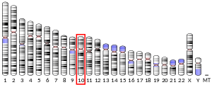
- ^ White RA, Dowler LL, Angeloni SV, Pasztor LM, MacArthur CA (November 1995). "Assignment of FGF8 to human chromosome 10q25-q26: mutations in FGF8 may be responsible for some types of acrocephalosyndactyly linked to this region". Genomics. 30 (1): 109–11. doi:10.1006/geno.1995.0020. PMID 8595889.
- ^ a b c "Entrez Gene: FGF8 fibroblast growth factor 8 (androgen-induced)".
- ^ Harris WA, Sanes DH, Reh TA (2011). Development of the Nervous System, Third Edition. Boston: Academic Press. pp. 33–34. ISBN 978-0-12-374539-2.
- ^ Crossley PH, Martin GR (February 1995). "The mouse Fgf8 gene encodes a family of polypeptides and is expressed in regions that direct outgrowth and patterning in the developing embryo". Development. 121 (2): 439–51. doi:10.1242/dev.121.2.439. PMID 7768185.
- ^ Grove EA, Fukuchi-Shimogori T (2003). "Generating the cerebral cortical area map". Annual Review of Neuroscience. 26: 355–80. doi:10.1146/annurev.neuro.26.041002.131137. PMID 14527269.
- ^ Rebsam A, Seif I, Gaspar P (October 2002). "Refinement of thalamocortical arbors and emergence of barrel domains in the primary somatosensory cortex: a study of normal and monoamine oxidase a knock-out mice". The Journal of Neuroscience. 22 (19): 8541–52. doi:10.1523/JNEUROSCI.22-19-08541.2002. PMC 6757778. PMID 12351728.
- ^ a b Lewandoski M, Sun X, Martin GR (December 2000). "Fgf8 signalling from the AER is essential for normal limb development". Nature Genetics. 26 (4): 460–3. doi:10.1038/82609. PMID 11101846. S2CID 28105181.
- ^ Moon AM, Capecchi MR (December 2000). "Fgf8 is required for outgrowth and patterning of the limbs". Nature Genetics. 26 (4): 455–9. doi:10.1038/82601. PMC 2001274. PMID 11101845.
- ^ Edom-Vovard F, Bonnin M, Duprez D (October 2001). "Fgf8 transcripts are located in tendons during embryonic chick limb development". Mechanisms of Development. 108 (1–2): 203–6. doi:10.1016/s0925-4773(01)00483-x. PMID 11578876. S2CID 16604609.
- ^ Crossley PH, Minowada G, MacArthur CA, Martin GR (January 1996). "Roles for FGF8 in the induction, initiation, and maintenance of chick limb development". Cell. 84 (1): 127–36. doi:10.1016/s0092-8674(00)80999-x. PMID 8548816. S2CID 14188382.
- ^ a b c Hao Y, Tang S, Yuan Y, Liu R, Chen Q (March 2019). "Roles of FGF8 subfamily in embryogenesis and oral‑maxillofacial diseases (Review)". International Journal of Oncology. 54 (3): 797–806. doi:10.3892/ijo.2019.4677. PMID 30628659.
- ^ Estienne A, Price CA (January 2018). "The fibroblast growth factor 8 family in the female reproductive tract". Reproduction. 155 (1): R53–R62. doi:10.1530/REP-17-0542. PMID 29269444.
Further reading
- Powers CJ, McLeskey SW, Wellstein A (September 2000). "Fibroblast growth factors, their receptors and signaling". Endocrine-Related Cancer. 7 (3): 165–97. CiteSeerX 10.1.1.323.4337. doi:10.1677/erc.0.0070165. PMID 11021964.
- Mattila MM, Härkönen PL (2007). "Role of fibroblast growth factor 8 in growth and progression of hormonal cancer". Cytokine & Growth Factor Reviews. 18 (3–4): 257–66. doi:10.1016/j.cytogfr.2007.04.010. PMID 17512240.
- Duester G (June 2007). "Retinoic acid regulation of the somitogenesis clock". Birth Defects Research. Part C, Embryo Today. 81 (2): 84–92. doi:10.1002/bdrc.20092. PMC 2235195. PMID 17600781.
- Tanaka A, Miyamoto K, Matsuo H, Matsumoto K, Yoshida H (April 1995). "Human androgen-induced growth factor in prostate and breast cancer cells: its molecular cloning and growth properties". FEBS Letters. 363 (3): 226–30. doi:10.1016/0014-5793(95)00324-3. PMID 7737407. S2CID 35818377.
- Gemel J, Gorry M, Ehrlich GD, MacArthur CA (July 1996). "Structure and sequence of human FGF8". Genomics. 35 (1): 253–7. doi:10.1006/geno.1996.0349. PMID 8661131.
- Ornitz DM, Xu J, Colvin JS, McEwen DG, MacArthur CA, Coulier F, et al. (June 1996). "Receptor specificity of the fibroblast growth factor family". The Journal of Biological Chemistry. 271 (25): 15292–7. doi:10.1074/jbc.271.25.15292. PMID 8663044.
- Payson RA, Wu J, Liu Y, Chiu IM (July 1996). "The human FGF-8 gene localizes on chromosome 10q24 and is subjected to induction by androgen in breast cancer cells". Oncogene. 13 (1): 47–53. PMID 8700553.
- Ghosh AK, Shankar DB, Shackleford GM, Wu K, T'Ang A, Miller GJ, et al. (October 1996). "Molecular cloning and characterization of human FGF8 alternative messenger RNA forms". Cell Growth & Differentiation. 7 (10): 1425–34. PMID 8891346.
- Yoshiura K, Leysens NJ, Chang J, Ward D, Murray JC, Muenke M (October 1997). "Genomic structure, sequence, and mapping of human FGF8 with no evidence for its role in craniosynostosis/limb defect syndromes". American Journal of Medical Genetics. 72 (3): 354–62. doi:10.1002/(SICI)1096-8628(19971031)72:3<354::AID-AJMG21>3.0.CO;2-R. PMID 9332670.
- Chellaiah A, Yuan W, Chellaiah M, Ornitz DM (December 1999). "Mapping ligand binding domains in chimeric fibroblast growth factor receptor molecules. Multiple regions determine ligand binding specificity". The Journal of Biological Chemistry. 274 (49): 34785–94. doi:10.1074/jbc.274.49.34785. PMID 10574949.
- Loo BB, Darwish KK, Vainikka SS, Saarikettu JJ, Vihko PP, Hermonen JJ, et al. (May 2000). "Production and characterization of the extracellular domain of recombinant human fibroblast growth factor receptor 4". The International Journal of Biochemistry & Cell Biology. 32 (5): 489–97. doi:10.1016/S1357-2725(99)00145-4. PMID 10736564.
- Xu J, Liu Z, Ornitz DM (May 2000). "Temporal and spatial gradients of Fgf8 and Fgf17 regulate proliferation and differentiation of midline cerebellar structures". Development. 127 (9): 1833–43. doi:10.1242/dev.127.9.1833. PMID 10751172.
- Tanaka S, Ueo H, Mafune K, Mori M, Wands JR, Sugimachi K (May 2001). "A novel isoform of human fibroblast growth factor 8 is induced by androgens and associated with progression of esophageal carcinoma". Digestive Diseases and Sciences. 46 (5): 1016–21. doi:10.1023/A:1010753826788. PMID 11341643. S2CID 30175286.
- Ruohola JK, Viitanen TP, Valve EM, Seppänen JA, Loponen NT, Keskitalo JJ, et al. (May 2001). "Enhanced invasion and tumor growth of fibroblast growth factor 8b-overexpressing MCF-7 human breast cancer cells". Cancer Research. 61 (10): 4229–37. PMID 11358849.
- Mattila MM, Ruohola JK, Valve EM, Tasanen MJ, Seppänen JA, Härkönen PL (May 2001). "FGF-8b increases angiogenic capacity and tumor growth of androgen-regulated S115 breast cancer cells". Oncogene. 20 (22): 2791–804. doi:10.1038/sj.onc.1204430. PMID 11420691. S2CID 22624526.
- Zammit C, Coope R, Gomm JJ, Shousha S, Johnston CL, Coombes RC (April 2002). "Fibroblast growth factor 8 is expressed at higher levels in lactating human breast and in breast cancer". British Journal of Cancer. 86 (7): 1097–103. doi:10.1038/sj.bjc.6600213. PMC 2364190. PMID 11953856.
- Brondani V, Klimkait T, Egly JM, Hamy F (June 2002). "Promoter of FGF8 reveals a unique regulation by unliganded RARalpha". Journal of Molecular Biology. 319 (3): 715–28. doi:10.1016/S0022-2836(02)00376-5. PMID 12054865.
- Gnanapragasam VJ, Robson CN, Neal DE, Leung HY (August 2002). "Regulation of FGF8 expression by the androgen receptor in human prostate cancer". Oncogene. 21 (33): 5069–80. doi:10.1038/sj.onc.1205663. PMID 12140757.
External links
- GeneReviews/NCBI/NIH/UW entry on Kallmann syndrome
- FGF8 human gene location in the UCSC Genome Browser.
- FGF8 human gene details in the UCSC Genome Browser.
- v
- t
- e
PDB gallery
-
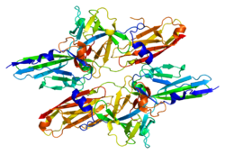
 2fdb: Crystal Structure of Fibroblast growth factor (FGF)8b in complex with FGF Receptor (FGFR) 2c


