Iliolumbar artery
| Iliolumbar artery | |
|---|---|
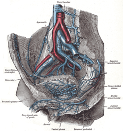 The veins of the right half of the male pelvis. (Iliolumbar artery not labeled, but Iliolumbar vein visible at center right.) | |
| Details | |
| Source | Internal iliac artery |
| Branches | Lumbar branches of iliolumbar artery |
| Vein | Iliolumbar vein |
| Supplies | Lumbar vertebrae, ilium |
| Identifiers | |
| Latin | arteria iliolumbalis |
| TA98 | A12.2.15.002 |
| TA2 | 4304 |
| FMA | 18845 |
| Anatomical terminology [edit on Wikidata] | |
The iliolumbar artery is the first branch of the posterior trunk of the internal iliac artery.
Structure
The iliolumbar artery is the first branch of the posterior trunk of the internal iliac artery.[1] It turns upward behind the obturator nerve and the external iliac artery and vein, to the medial border of the psoas major muscle, behind which it divides into:
Anastomoses
- 1. Last lumbar→iliolumbar
- 2. Lateral sacral↔lateral sacral
- 3. Middle sacral→lateral sacral
- 4. Superior hemorrhoidal→middle hemorrhoidal
- 5. Medial femoral circumflex→inferior gluteal
- 6. Medial femoral circumflex↔obturator
- 7. Lateral femoral circumflex→superior gluteal
- 8. Deep iliac circumflex→superior gluteal
- 9. Deep iliac circumflex→external iliac
- 10. Last lumbar→superior gluteal
- 11. Last lumbar→deep iliac circumflex
- 12. Iliolumbar→deep iliac circumflex.[2]
Additional images
-
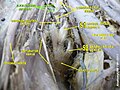 Iliolumbar artery

References
![]() This article incorporates text in the public domain from page 621 of the 20th edition of Gray's Anatomy (1918)
This article incorporates text in the public domain from page 621 of the 20th edition of Gray's Anatomy (1918)
- ^ Paterson-Brown, Sara (2010-01-01), Bennett, Phillip; Williamson, Catherine (eds.), "Chapter Five - Applied anatomy", Basic Science in Obstetrics and Gynaecology (Fourth Edition), Churchill Livingstone, pp. 57–95, doi:10.1016/b978-0-443-10281-3.00009-9, ISBN 978-0-443-10281-3, retrieved 2021-02-07
- ^ Chait, A; Moltz, A; Nelson, JH (February 1968). "The collateral arterial circulation in the pelvis. An angiographic study". American Journal of Roentgenology. 102 (2): 392–400. doi:10.2214/ajr.102.2.392. PMID 5635691.
External links
- Anatomy photo:44:10-0100 at the SUNY Downstate Medical Center
- Radiology image: Pelvis:15PelArt from Radiology Atlas at SUNY Downstate Medical Center (need to enable Java)
- Anatomy figure: 43:07-02 at Human Anatomy Online, SUNY Downstate Medical Center
- pelvis at The Anatomy Lesson by Wesley Norman (Georgetown University) (pelvicarteries)
- Illustration at mrcog-wiseowl.com
- v
- t
- e
Arteries of the abdomen and pelvis
aorta
| Inferior phrenic | |||||||||||||||||||||||||
|---|---|---|---|---|---|---|---|---|---|---|---|---|---|---|---|---|---|---|---|---|---|---|---|---|---|
| Celiac |
| ||||||||||||||||||||||||
| Superior mesenteric | |||||||||||||||||||||||||
| Suprarenal | |||||||||||||||||||||||||
| Renal | |||||||||||||||||||||||||
| Gonadal | |||||||||||||||||||||||||
| Lumbar | |||||||||||||||||||||||||
| Inferior mesenteric | |||||||||||||||||||||||||
| Common iliac |
| ||||||||||||||||||||||||
| Median sacral | |||||||||||||||||||||||||
Portal:
 Anatomy

| Authority control databases |
|
|---|
 | This cardiovascular system article is a stub. You can help Wikipedia by expanding it. |
- v
- t
- e


