Optic recess
| Optic recess | |
|---|---|
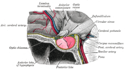 The hypophysis cerebri, in position. Shown in sagittal section. (Optic recess labeled at upper right.) | |
 Median sagittal section of brain. The relations of the pia mater are indicated by the red color. (Optic recess labeled at lower left.) | |
| Details | |
| Identifiers | |
| Latin | recessus supraopticus |
| NeuroNames | 457 |
| NeuroLex ID | nlx_144280 |
| TA98 | A14.1.08.418 |
| TA2 | 5773 |
| FMA | 78455 |
| Anatomical terms of neuroanatomy [edit on Wikidata] | |
At the junction of the floor and anterior wall of the third ventricle, immediately above the optic chiasma, the ventricle presents a small angular recess or diverticulum, the optic recess (or supraoptic recess).
Additional images
-
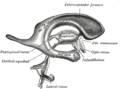 Drawing of a cast of the ventricular cavities, viewed from the side.

References
![]() This article incorporates text in the public domain from page 816 of the 20th edition of Gray's Anatomy (1918)
This article incorporates text in the public domain from page 816 of the 20th edition of Gray's Anatomy (1918)
- v
- t
- e
Ventricular system of the human brain
- Body
- Lamina affixa
- Stria terminalis
- Collateral eminence
- Occipital horn
- Calcar avis
- Septum pellucidum
| Roof | |
|---|---|
| Floor | |
| Apertures | |
| Other |
- Blood–brain barrier
- Cerebral aqueduct
- Interventricular foramina
- Perilymphatic duct
Portal:
 Anatomy

| Authority control databases |
|
|---|
 | This neuroanatomy article is a stub. You can help Wikipedia by expanding it. |
- v
- t
- e


