Palatine nerves
| Palatine nerves | |
|---|---|
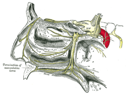 The sphenopalatine ganglion and its branches. (Anterior palatine at bottom right, middle palatine at bottom center, and posterior palatine at bottom right.) | |
| Details | |
| Identifiers | |
| Latin | nervi palatini |
| Anatomical terms of neuroanatomy [edit on Wikidata] | |
The palatine nerves (descending branches) are distributed to the roof of the mouth, soft palate, tonsil, and lining membrane of the nasal cavity.
Most of their fibers are derived from the sphenopalatine branches of the maxillary nerve.
In older texts, they are usually categorized as three in number: anterior, middle, and posterior. (In newer texts, and in Terminologia anatomica, they are broken down into "greater palatine nerve" and "lesser palatine nerve".)
References
![]() This article incorporates text in the public domain from page 893 of the 20th edition of Gray's Anatomy (1918)
This article incorporates text in the public domain from page 893 of the 20th edition of Gray's Anatomy (1918)
External links
- lesson9 at The Anatomy Lesson by Wesley Norman (Georgetown University)
- MedEd at Loyola GrossAnatomy/h_n/cn/cn1/cnb2.htm
- Diagram at adi-visuals.com
- v
- t
- e
The cranial nerves
- Nuclei
- septal nuclei
- Course
- no significant branches
- Nuclei
- anterior olfactory nucleus
- Course
- Nuclei
- Course
- Nuclei
- Branches
- Nucleus
- Branches
- no significant branches
- Nuclei
- Course
- Branches
- Nucleus
- Branches
- no significant branches
| Near origin | |
|---|---|
| Inside facial canal | |
| At stylomastoid foramen | |
| Nuclei |
| Before jugular fossa | |
|---|---|
| After jugular fossa | |
| Nuclei |
| Before jugular fossa | |
|---|---|
| After jugular fossa | |
| Neck | |
| Thorax |
|
| Abdomen | |
| Nuclei |
 | This neuroanatomy article is a stub. You can help Wikipedia by expanding it. |
- v
- t
- e
Portal:
 Anatomy


