Thùy chẩm là một trong bốn thùy chính của vỏ đại não trong não của động vật có vú. Thùy chẩm là trung tâm xử lý thị giác của não động vật có vú, chứa hầu hết các vùng giải phẫu của vỏ thị giác.[1] Vỏ não thị giác chính là khu vực Brodmann 17, thường được gọi là V1 (visual one). V1 của người nằm ở phía giữa của thùy chẩm bên trong rãnh cựa; phạm vi đầy đủ của V1 thường tiếp tục tới cực sau của thùy chẩm. V1 thường được gọi là vỏ não có sọc bởi vì có thể nhận diện nó bằng một đường sọc myelin lớn Stria of Gennari.
Cấu trúc
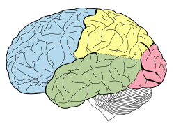
Hoạt họa. Thùy chẩm (đỏ) của bán cầu đại não trái.
Hai thùy chẩm là cặp thùy nhỏ nhất trong số bốn cặp thùy của vỏ đại não người. Nằm ở vị trí tận cùng của sọ, thùy chẩm là một phần của não trước. Không một thùy thuộc vỏ não nào được định nghĩa bằng bất cứ đặc điểm cấu trúc bên trong nào, mà thay vào đó bởi các xương của xương đầu mà nằm phía trên chúng. Do đó, thùy chẩm được định nghĩa như một phần của vỏ đại não nằm bên dưới xương chẩm. (Xem bài não người để biết thêm chi tiết.)
Tổn thương đến các khu vực thị giác chính của thùy chẩm có thể khiến người đó bị mù một phần hoặc hoàn toàn.[2]
Chức năng
Một khía cạnh về mặt chức năng quan trọng của thùy chẩm là nó chứa đựng vỏ thị giác chính.
Hình ảnh
Base of brain.
Drawing to illustrate the relations of the brain to the skull.
Occipital lobe in blue
Thùy chẩm
Thùy chẩm
Ventricles of brain and basal ganglia.Superior view. Horizontal section.Deep dissection
Tham khảo
^“SparkNotes: Brain Anatomy: Parietal and Occipital Lobes”. Bản gốc lưu trữ ngày 31 tháng 12 năm 2007. Truy cập ngày 27 tháng 2 năm 2008.
^Schacter, D. L., Gilbert, D. L. & Wegner, D. M. (2009). Psychology. (2nd ed.). New Work (NY): Worth Publishers.


 Base of brain.
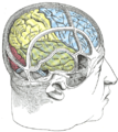 Drawing to illustrate the relations of the brain to the skull.
 Occipital lobe in blue
 Thùy chẩm
 Thùy chẩm
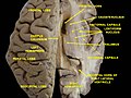 Ventricles of brain and basal ganglia.Superior view. Horizontal section.Deep dissection


